

Need Immediate Help? Call Toll Free 800.866.6689
The Sequoia system represents the most advanced technology in ultrasound imaging. And each unique technology and innovation is designed to help you make more confident and accurate diagnoses, delivering excellent image quality and enhancing workflow, across a broad range of patient types and applications.
The Sequoia system is also engineered to help make your practice as efficient as possible. It is designed to be the centerpiece of your diagnostic or research practice, today, tomorrow and far into the future: a comprehensive, state-of-the-art ultrasound system without equal and without compromise.
The ACUSON Sequoia ultrasound system is synonymous with unparalleled imaging performance, patient after patient, in a wide range of clinical applications. Siemens continues to lay the foundation for the future of ultrasound, reaffirming our commitment to imaging excellence and unceasing innovation that will continue to put the Sequoia system at the forefront of ultrasound performance into the 21st century.
- TEQ Ultrasound Technology
- ▣ TEQ ultrasound technology for 2D is a sophisticated signal-processing technology that automatically equalizes tissue gain and brightness in two dimensions providing consistent, reproducible image quality in 2D and M-mode at the push of a button
- ▣ TEQ ultrasound technology for spectral Doppler adapts to individual patient hemodynamics and instantly optimizes for PW and CW Doppler information, saving time and maximizing workflow- Automatically optimizes scale, baseline, gain and dynamic range at the push of a button
- ▣ Pre-processing technology applied to RF echo data before image is formed
- ▣ Affords increased productivity and reduced inter-operator variability
- ▣ Overcomes limitations of conventional DGC adjustments for non-uniform tissue structures
- ▣ Assures optimal gain settings under suboptimal ambient lighting conditions for stored images
- ▣ Assists in image brightness settings during contrast agent imaging studies
- ▣ Frees operator to focus on imaging, not on image quality adjustments
- ▣ Available on all transducers
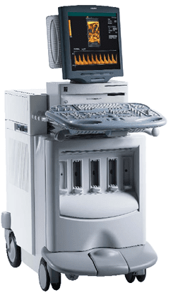
- Monitor
- ▣ 14" (36 cm) high-resolution non-interlaced computer color monitor; 11.8" (30 cm) viewable screen size
- ▣ Resolution:
- - PAL: 768X576, 50 Hz
- - NTSC: 640X480, 60 Hz
- Operating/Display Modes
- ▣ 2D
- ▣ Native tissue harmonic imaging
- ▣ Transmit Compounding
- ▣ Spatial Compounding
- ▣ Spatial Compounding Plus
- ▣ SST color Doppler (CD) Imaging including:
- - CDV Color Doppler Velocity capability
- - CDE Color Doppler Energy capability
- - Convergent color Doppler capability (CCD color)
- - DTI Doppler tissue imaging capability
- - DTI Harmonics
- - Color Harmonic Imaging capability
- ▣ Solo spectral Doppler imaging including:
- - PW (Pulsed Wave) and HPRF (High Pulse Repetition Frequency) Spectral
- Doppler modes
- - DT (Doppler Tissue) PW Spectral Doppler mode
- - CW (Continuous Wave) Spectral Doppler mode
- ▣ M-mode/Color Doppler M-mode
- ▣ Cadence contrast agent imaging technology
- - Cadence contrast pulse sequencing technology (CPS) for Cardiology and General Imaging
- - Cadence agent detection imaging technology (ADI) for General Imaging
- - Cadence power contrast imaging (PCI) for Cardiology
- - Cadence coherent contrast imaging technology (CCI) for Cardiology
- ▣ Supported combinations of modes:
- - 2D
- - 2D/M-mode
- - M-mode
- - 2D/PW or HPRF (simultaneous or update)
- - PW or HPRF
- - 2D/CW, simultaneous or update (for Cardiac Option)
- - CW (for Cardiac Option)
- - 2D/CDV
- - 2D/CDV/PW or HPRF
- - 2D/CDV/CW (for Cardiac Option)
- - 2D/CDE
- - 2D/CDE/PW or HPRF
- - 2D/CCD
- - 2D/CCD/PW or HPRF
- - 2D/DTI
- - 2D/DTI/DTPW
- - 2D/CPS
- - 2D/PCI
- - 2D/DTI/Color Doppler M-mode
- - 2D/CDV/Color Doppler M-mode Dual Screen and Live Dual Imaging
- 2D Calipers - Generic Measurements and Calculations
- ▣ 6 pairs of 2D calipers available.
- Screen display:
- - Depth for single calipers
- - Distance between calipers for each pair
- ▣ Ellipse function: Up to 6 pairs of calipers.
- Displays for each pair of calipers include:
- - Distance between calipers
- - Ellipse circumference
- - Ellipse area
- ▣ Trace function. Displays:
- - Caliper depth
- - Trace circumference
- - Traced area
- ▣ Minimum distance between calipers:
- - Transducer type, depth and RES enhanced resolution setting dependent
- - 0.003 cm
- Cine Capability
- ▣ Available in all modes
- ▣ Imaging Cine, for real-time acquisition and review of 2D and color Doppler images
- ▣ Strip Cine, for real-time acquisition and review of spectral Doppler or M-mode strips in full strip or combined screen
- ▣ Continuous acquisition
- ▣ Dedicated "push-rotate" wheel control for freezing and immediate scrolling through Cine memory
- ▣ Adjustable memory allocated for Cine (from 20 to 80% of available RAM)
- ▣ Number of frames or seconds of information in Cine memory depends on:
- - Mode in use
- - Image adjustment
- - Amount of information displayed (2D, color Doppler, image size, image texture, etc.)
- - Adjustable memory allocated for Cine
- ▣ Measurement and calculation capability Imaging Cine
- The DIMAQ Integrated Ultrasound Workstation Integral to Sequoia 512 system architecture is a special purpose DIMAQ workstation. The DIMAQ workstation offers advanced digital image and data processing and management in real time. It has direct access to all information in the ultrasound system for the current and previous exams.
- ▣ Real-time digital image processing, such as DELTA differential echo amplification
- ▣ Exam Categories, Exam Presets, Image Presets configurations
- ▣ Digital image and data management system
- ▣ Hard drive: 18GB
- ▣ RAM size: 64 MB
- ▣ 640 Mb magneto-optical disc drive compatible with 3-1/2" MO discs of 128, 230 and 540 Mb capacity
- ▣ Digital capture, storage and retrieval of images and data:
- - Review capability of stored examination from the hard disc or MO disc for review or calculations